Primary T abnormalities (Incomplete)
Myocardial ischemia / infarction, pericarditis, myocarditis, CNS, apical HOCM, medication effect. Other less common conditions include advanced AV block, S/P carotid endarterectomy, pheochromocytoma, cocaine abuse, cardiac metastases. |
T
wave inversion | ST
| |||
Symmetrical | Asymmetrical | Peak | Round | |
Yes | . | Yes | . | Usually
no siginificant deviation |
| Patient, 48 year old male was seen in ER an hour after onset of chest pain. ECG on arrival revealed anterior MI. He underwent an urgent primary PCI and stent placement of the near occluded LAD lesion. Echocardiogram showed only focal area of akinesis involving distal septum and apex with preserved overall systolic function. First ECG was performed on 5/24/04 at 9.56 am (Not shown here). The subsequent ECGs were done following the PCI procedure (Shown below) | |||||
| Example of primary T wave abnormalities in V3-V4 and I. V5-V6 has characteristic in between primary and secondary T wave abnormalities. | |||||
5/25/04. | I
 | V3 | V4 | V5 | V6 |
| Example of giant T wave inversion | |||||
5/26/04. | I
 | V3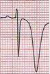 | V4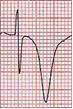 | V5 | V6 |
| Go
2ry T wave abnormalities Go to 1ry and 2ry T abnormalities in one patient |