 |
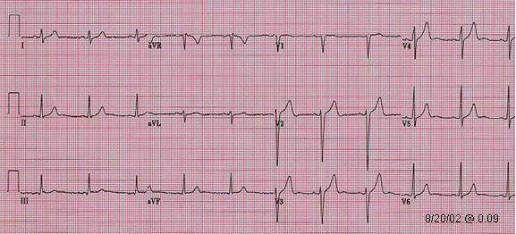
| 5 hours before acute anterior MI with hyperacute T wave pattern |
| Patient. 50 year old male had recurrent anterior chest pain radiating to left shoulder and arm few hours before admission. First ECG in ER was at 23.27 of 10/19/02 which is identical to the one above. He had no chest pain and troponin was negative at that time. Five hours later he had severe chest pain again and ECG was repeated (below). The patient received an emergency direct infarct PCI/stent procedure to the complete occluded proximal LAD. Subsequent troponin were elevated. ECG on the next page show evolving of ST elevation anterior MI pattern following reperfusion treatment. |
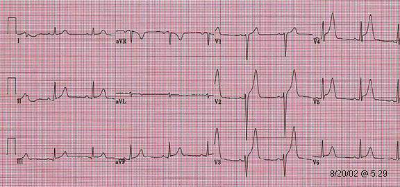 |
| Acute anterior MI. Hyperacute T wave |
Note: (1). ST T change in this case also look similar to pericarditis or early repolarization pattern by itself. (2). No reciprocal change. (3). ST elevation in both anterior and inferior leads may represent injury involving both anterior, inferior wall and wrap around the apex. |
Go to further evolving MI pattern of this patient with pseudonormalization
Go
to ST T abnormalities in acute ST elevation MI table
Go
to Evolving ST T abnormalities in acute ST elevation MI
Go
to anterior MI menu /
Go to MI menu /
Go to main menu