 |
I |
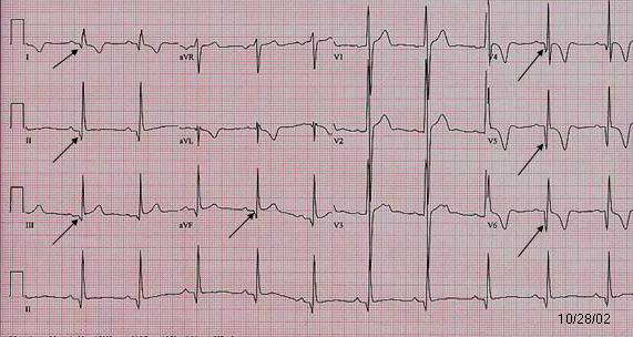
| HOCM. (Case 2) Producing pseudo Q wave |
| HOCM (Hypertrophic Obstructive CardioMyopathy) usually has LVH pattern. High R wave in V1-V3 in this case may be due to septal hypertrophy. |
| Patient. 25 year old male was diagnosed having HOCM by echocardiogram since he was 18 year old during a routine visit for an upper respiratory infection and a murmur was heard. Most recent echocardiogram continue to show typical pattern of a HOCM with systolic anterior motion of the mitral valve anterior leaflet, mild to moderate LV outflow tract obstruction and mild to moderate mitral regurgitation. The left atrium is slightly enlarged. |
 II
II III
III V4
V4 V5
V5 V6
V6
Go
to Giant T wave inversion menu /
Go
to pseudo Q wave
Go
to LVH menu / Go
to HOCM menu / Go
to specific diagnosis / Go
to main menu