 |
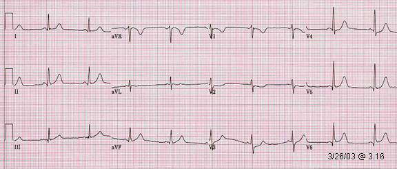
| Infero-posterior MI (ECG 1) |
| Patient. 54 year old male with previous inferior MI and underwent PCI/stent placement to circumflex and obtuse marginal branches in 7/02. Brachytherapy was performed for instent restenosis in these areas in 11/02. He was brought in this time with severe chest pain and became extremely agitate required heavy sedation and intubation during emergent PCI/drug eluded stent to circumflex and obtuse marginal branches again. |
| Comment. ST elevation in II, III, aVF, V6 is minimum, if there is any at all. Reciprocal ST depression in V1-3 is more easily recognized. The ST depression in V1-3 may lead to the diagnosis of non Q wave anterior MI and immediate reperfusion therapy will not be considered. |
| . |
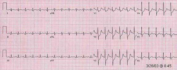 |
| Infero-posterior MI (ECG 2) |
| Further ECG changes: (1). Prominent R wave in early precordial leads (R/S >1). (2). Cannot exclude development of incomplete RBBB which may also cause prominent R in V1. (3). More ST depression in V1-4. 1# and 3# is consistent with acute posterior MI. ST T pattern in II, III, aVF, I and aVL is suspicious for diagnosis of infero-lateral MI. |
Go
to ECG 3 of the same patient
Go
to prominent
R in V1 (R/S >1)
Go
to MI menu
Go to main
menu