 |
II V1 V1 V6 V6 |
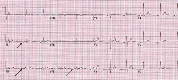
| Ectopic atrial rhythm (Case 1) |
| Patient, 70 year old male who was admitted with abdominal pain, nausea, vomiting, and diarrhea. He subsequently underwent successful laparoscopic cholecystectomy 2 days later. Other diagnosis are hypertension, hyperlipidemia, and S/P abdominal aortic aneurysm repair. Echocardiogram showed left atrial enlargement of 46 mm, concentric LVH with normal size and systolic function. Right heart size and calculated pulmonary artery pressure were normal. Other ECGs showed normal P wave. |
Criteria |
| There are wide range in variation of sinus P wave since several factors may effect the P wave. These include: Anatomic cardiac position to the anterior chest wall; state of autonomic influence; intra and/or interatrial conduction abnormality; pressure and/or volume load; atrial/atria diseases; electrode positioning (precordial lead). |